What to Expect
It can be stressful to have a radiology procedure, particularly the first time. To help you know what to expect, here’s a list of our procedures and their descriptions. Just click on a procedure name to find out more.
Bone Densitometry (DXA)
 Measurement of bone mass by densitometry has become central to the diagnosis of osteoporosis and instituting preventative treatment. Currently it is estimated that less than 5% of patients at risk for osteoporotic fractures of the spine and hip are diagnosed and treated. With increased availability of accurate bone density testing, diagnosis of osteoporosis early will allow for earlier treatment, resulting in reduced fracture risk. There are different testing techniques to predict future fracture risk. However, only central measurements of the hip and spine can confirm the diagnosis of osteoporosis, determine the severity of the osteoporosis, decide when therapy should be initiated, and monitor response to therapy.
Dual-Energy X-Ray Absorptometry (DXA) units send a small column of x-ray to a detector. The units are state-of-the-art, providing the highest levels of accuracy in bone mineral determination. The test results are stored in the computer to allow direct comparison to future studies.
Indications to Refer Patients for Bone Densitometry Testing Include:
- Diagnose osteoporosis-even in its earliest stages
- Estimate risk of future fractures
- Monitor efficacy of drug therapy on bone density
- Evaluate children for suspected low bone mass
What to Expect:
A comprehensive questionnaire will be given prior to the exam to identify your risk factors. We try to identify all risk factors, since the presence of multiple risk factors may require that you be treated earlier or tested more frequently. We calculate a FRAX score for postmenopausal women or older men who are in the osteopenic range to determine if they are also candidates for treatment.
DXA scanning is a simple, painless test, performed while lying down on the padded table of the unit. Measurements are made in the lumbar spine, hip and occasionally the forearm. In pediatric patients, measurements are only made in the lumbar spine. There is no injection. The length of the exam is 10-15 minutes.
Preparation:
- No calcium supplements, day of exam.
- Please bring a list of all medications to your appointment.
- Wear clothing without metallic fasteners or decorations, if you do not wish to change into a hospital gown.
You should not have a barium study, radioisotope injection, or oral or IV contrast material from a CT scan within 7 days prior to the DXA scan. You cannot have this exam if you are pregnant, but it is safe if you are nursing.
Locations for Bone Densitometry:
- St. Francis Medical Center
- St. Mary’s Hospital
- Bon Secours Imaging at Innsbrook
- Bon Secours Imaging at Reynolds
- Bon Secours Imaging at Westchester
- Laburnum Diagnostic Imaging Center
Breast MRI
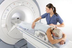 Magnetic Resonance Imaging (MRI) of the breast – or breast MRI – is a non-invasive test that assists in diagnosing problems in the breast by using a strong magnetic field. MRI uses a combination of magnets and radiowaves to test the breast. The scanner then receives the natural radio frequencies emitted by tissues in the breast to create images interpreted by a radiologist.
Mammograms use x-rays to generate images of the breast tissue to search for cancer. Breast MRI is a different imaging technique that captures multiple cross-sectional pictures of your breast without any ionizing radiation exposure. Breast MRI looks at the blood supply to the breast. Breast MRI does not replace a mammogram, but is an additional screening tool used for women at high-risk of developing breast cancer.
Women with a greater than 20% lifetime risk for developing breast cancer should consider being screened using breast MRI, along with their annual mammogram. Women are considered high-risk if they have a strong family history of breast or ovarian cancer, received chest radiation for Hodgkin’s disease, or have a genetic predisposition for breast cancer. Breast MRI may also be recommended for women who have an increased risk of breast cancer.
Breast MRI poses minimal risk to most patients. However, since the MRI scanner uses a strong magnet and radio waves to create images, there may be a danger in screening patients who have implanted mechanical devices such as pacemakers that use electricity to operate. Before the scan, the patient will be asked to fill out a detailed questionnaire to determine if any risks are involved. There will be additional questions about prior history of breast cancer, prior high-risk benign biopsy, or dense breast tissue. Your doctor can help you determine your personal risk level and if breast MRI is appropriate for you.
What to Expect:
During the MRI scan, you will lie face down on your stomach. There is no compression of the breasts during the exam. There is no special preparation. Contrast is used in this exam to help highlight any breast abnormalities. The contrast is injected intravenously during the exam and is hypoallergenic. The clear contrast passes out of the body in about a day. The duration of the exam is about 45 minutes to 1 hour. The actual scan time will be about 30 minutes. Although MRI exams are covered by insurance, please contact your insurance company for your personal coverage guidelines.
Locations for Breast MRI:
- Memorial Regional Medical Center
- St. Francis Medical Center
- St. Mary’s Hospital
- Bon Secours Imaging Center at Reynolds Crossing
- Bon Secours Imaging at Westchester
Computed Tomography (CT)
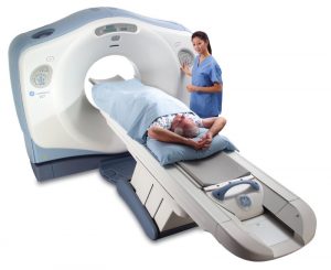 Computerized Tomography (CT) scans are special x-ray tests that produce cross-sectional images of the body using x-rays and a computer. These images allow the radiologist to look at the inside of the body just as one would look at the inside of a loaf of bread by slicing it. In most exams, two-dimensional or three-dimensional reconstructions are created for a comprehensive evaluation of the area. CT scans are frequently used to evaluate the brain, neck, spine, chest, abdomen, pelvis and sinuses. Sometimes, physicians will order special thin section CT scans to upload onto the interactive computers in the operating room during complex neurosurgery or ENT procedures or as a map to plan radiation therapy.
Head or brain CT evaluates the various structures of the brain to look for a mass, stroke, bleed or blood vessel abnormality. It is also sometimes used to evaluate the skull.
Neck CT evaluates the soft tissues of the neck and is frequently used to study a lump or mass in the neck or to look for enlarged lymph nodes or glands.
CT of the chest is frequently used to further evaluate an abnormality on a plain chest xray. It is also often used to evaluate the lungs or look for pulmonary embolism (blood clot) in the lungs or enlarged lymph nodes.
Abdominal and pelvic CT evaluates the abdominal and pelvic organs and the gastrointestinal tract. These studies are often ordered to evaluate for a cause of pain and sometimes to follow-up on an abnormality seen on another test such as an ultrasound.
Extremity CT exams are usually reconstructed into two-dimensional or three-dimensional images to allow the radiologist and orthopedist to fully understand the extent of a fracture or bone lesion and plan surgery.
Sinus CT exam is used to both diagnose sinus disease and to look for a narrowing or obstruction in the sinus drainage pathway.
Maxillofacial CT exam is used to understand complex fractures of the face.
Spine CT test is most commonly used to look for a herniated disc or narrowing of the spinal canal (spinal stenosis) in patients with neck, arm back and/or leg pain.
What to Expect:
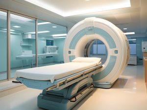
The CT scanner looks like a large donut with a narrow table. The patient lies on the table, which then slides into the center of the machine. The technologist is in the next room and can observe the patient through a large window. The x-ray tube moves rapidly around the area of anatomic interest. The sensor then sends the information to the computer. The computer reconstructs this data with special algorithms that are most sensitive to “see” findings in the bone, lung, brain, and soft tissues. Some scans (abdomen and pelvis) require the patient to drink a dilute barium liquid prior to the scan. Also, for certain exams contrast or dye must be injected into a vein during the scan. The entire procedure takes 15 to 45 minutes depending on what part of the body is being scanned. After the scan is finished, a board certified radiologist will interpret the study and send a report to the referring physician.
Preparation:
If you have ever had a prior allergic reaction to a contrast agent, we will need to know the type of contrast and the details of the reaction. We also need to know if you have a history of multiple allergies or asthma. Please let us know this information before your visit so that we can avoid the need to reschedule your appointment. We will be able to tell you if you will need premedication prior to your exam. If you are over age 65 or have diabetes, high blood pressure or any history of kidney or severe liver disease or transplant we will need the results of blood tests for kidney function before we give intravenous contrast. If you are a diabetic on metformin, you will be asked to stop taking your medication for 48 hours after your exam. A special instruction sheet will be given to you at the time of the exam.
Abdomen/Pelvis (renal stone protocol)
- No preparation necessary
Abdomen/ Pelvis
- Pick up one bottle of CT barium Scan C the day before your exam or arrive 2 hours prior to your exam
- Do not eat or drink anything except water for 4 hours before your exam.
- Two hours before your exam drink the whole bottle of barium (Scan C).
Instructions for oral barium:
- Scan C is pre-mixed and ready to use.
- It may taste better when refrigerated.
- Please SHAKE WELL before using.
- DO NOT mix with any other substance.
- DO NOT pour over ice.
- DO NOT have any liquid or solid food for FOUR HOURS prior to your scheduled appointment time.
- NOTE: During this preparation time, be sure to take all medications as prescribed by your physician.
Head/Neck/Chest/Extremities
- DO NOT eat or drink anything except water for 4 hours before your exam.
Sinus CT
- No preparation necessary
Locations for CT Exam:
- Memorial Regional Medical Center
- Richmond Community Hospital
- St. Francis Medical Center
- St. Mary’s Hospital
- Bon Secours Imaging at Innsbrook
- Bon Secours Imaging at Short Pump
- Bon Secours Imaging at Westchester
Diagnostic Mammogram and Breast Ultrasound
 A diagnostic mammogram is performed on women who have a breast problem or symptom, such as a lump, nipple discharge or breast pain or as a follow up to an abnormal screening mammogram.
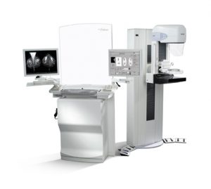
Diagnostic Mammography
Digital mammography, also called full-field digital mammography (FFDM), is a mammography system in which the x-ray film is replaced by solid-state detectors that convert x-rays into electrical signals. These detectors are similar to those found in digital cameras. The electrical signals are used to produce images of the breast that can be seen on a computer screen or printed on special film similar to conventional mammograms. From the patient’s point of view, digital mammography is essentially the same as the screen-film system.
Computer-aided detection (CAD) systems are used, when possible, as essentially a “second read” of our digitally acquired mammogram. The computer software then searches for abnormal areas of density, mass, or calcification that may indicate the presence of cancer. The CAD system highlights these areas on the images, alerting the radiologist to the need for further analysis.
3D mammography (also called Tomosynthesis) can be requested at some of our facilities. The camera moves in an arc over the breast, and then the computer reconstructs the images into a cine loop. The 3D image set helps distinguish masses within dense tissue and minimizes the tissue overlap that can hide cancers.
Breast Ultrasound
 Breast ultrasound is performed to evaluate a palpable lump or an abnormality visualized on a mammogram. The distinction can be made between cystic (fluid-filled) and solid abnormalities. This information aides the radiologist and the patient’s clinician in determining the appropriate course of treatment for the patient. The exam is performed by a registered sonographer and interpreted by a Board Certified radiologist that both specialize in breast imaging.
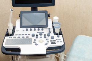
Ultrasound uses high frequency sound waves instead of radiation to image the breast tissue. The exam is performed by placing a small amount of warm gel on the breast, then scanning the area of concern with an ultrasound transducer.
What to Expect:
The radiographs taken vary and are dedicated to the particular finding or problem. The mammogram is performed on a dedicated x-ray machine that is designed to provide the best possible pictures with the least amount of radiation. To achieve this, the breast(s) is/are compressed briefly during the exam. The length of the exam varies. Depending on the findings of the diagnostic mammogram, a breast ultrasound may follow. The diagnostic mammogram is performed by a technologist who has advanced training and certification in breast imaging. After the x-rays are obtained, they are reviewed and interpreted by a board certified radiologist, who specializes in breast imaging. All facilities are accredited in mammography by The American College of Radiology and are inspected yearly by the FDA.
All diagnostic mammograms are performed with a radiologist who specializes in breast imaging on the premises. The radiologist will be available to answer any questions you may have.
It is important for the radiologist to compare your current films with any prior films you may have had. If your previous mammogram was not performed at the facility where you have your exam, please bring the films with you to your appointment or tell the scheduler where they were taken, so that we can obtain them in time for your appointment.
Preparation:
Do not wear any powder or deodorant under the arms or around the breast area.
Locations for Diagnostic Mammography:
- Memorial Regional Medical Center
- St. Mary’s Hospital
- Bon Secours Imaging at Westchester
Mammogram (Screening)
A screening mammogram is performed on women with no symptoms to detect abnormalities of the breasts. Mammography uses a low dose radiographic system with a high contrast, high resolution detector or film. Mammography has been shown to detect 85-90% of breast cancer in women over the age of 50. Current guidelines recommend baseline mammography, then annual mammography, beginning at age 40. A patient who has had a first degree relative (mother, sister, or daughter) with breast cancer should start annual screening 10 years prior to the age of their relative’s diagnosis.
Digital mammography, also called full-field digital mammography (FFDM), is a mammography system in which the x-ray film is replaced by solid-state detectors that convert x-rays into electrical signals. These detectors are similar to those found in digital cameras. The electrical signals are used to produce images of the breast that can be seen on a computer screen or printed on special film similar to conventional mammograms. From the patient’s point of view, digital mammography is essentially the same as the screen-film system.
Computer-aided detection (CAD) systems are used, when possible, as essentially a “second read” of our digitally acquired mammogram. The computer software then searches for abnormal areas of density, mass, or calcification that may indicate the presence of cancer. The CAD system highlights these areas on the images, alerting the radiologist to the need for further analysis.
3D mammography (also called Tomosynthesis) can be requested at some of our facilities. The camera moves in an arc over the breast, and then the computer reconstructs the images into a cine loop. The 3D image set helps distinguish masses within dense tissue and minimizes the tissue overlap that can hide cancers.
What to Expect:
Four x-ray pictures, two of each breast are taken on a dedicated x-ray machine. To obtain the best possible images with the least radiation the breasts are compressed briefly. At all facilities, we use a disposable cushion (Mammopad) between the breast and the machine to make the procedure more comfortable. The exam takes approximately one half hour. The mammogram is performed by a female technologist who has advanced training and certification in breast imaging. No radiologist is present during the study for a screening exam; the test is commonly reviewed and interpreted the following day by a board certified radiologist, who specializes in breast imaging. All facilities are accredited in mammography by The American College of Radiology and are inspected yearly by the FDA.
It is not uncommon for a radiologist to ask the technologist to perform special or additional x-ray pictures or a breast ultrasound to see an area better. Additional images are not always the sign of a serious problem. The interpretation of the films will be sent to your doctor and you will receive a letter with the results of your mammogram. At the time of service, you can ask that a normal result be sent to you by email; any result that requires a short term follow-up, 6 month follow-up or biopsy will be sent to you by US mail.
It is important for the radiologist to compare your current mammogram with your prior mammogram. If your prior films were not obtained at the facility where you have your exam, please bring them with you to your appointment or tell the scheduler where they were taken, so that we may obtain them in time for your appointment.
Preparation:
Do not wear any powder or deodorant under the arms or around the breast area.
Locations for Screening Mammography:
- Memorial Regional Medical Center
- Richmond Community Hospital
- St. Francis Medical Center
- St. Mary’s Hospital
- Bon Secours Imaging at Innsbrook
- Bon Secours Imaging at Reynolds Crossing
- Bon Secours Imaging at Short Pump
- Bon Secours Imaging at Westchester
- Laburnum Diagnostic Imaging Center
Magnetic Resonance Imaging (MRI)
 Magnetic Resonance Imaging (MRI) uses a strong magnetic field and radiowaves to image the body. MRI is commonly used to examine the brain, spine, joints, abdomen and pelvis. A special kind of MRI exam, called magnetic resonance angiography (MRA), examines the blood vessels.
Brain MRI
An MRI of the brain produces detailed pictures of the brain. It is commonly used to study patients with headaches, seizures, weakness, blurry vision, etc. It also can further evaluate an abnormality seen on a CT scan. During the brain MRI, a special device called a head coil is placed around the patient’s head. It does not touch the patient, and the patient can see through large gaps in the coil. This device is what helps to produce the detailed pictures of the brain.
Spine MRI
This test is most commonly used to look for a herniated disc or narrowing of the spinal canal (spinal stenosis) in patients with neck, arm back and/or leg pain. It is also the best test to look for a recurrent disc herniation in a patient who has had prior back surgery.
Bone and Joint MRI
MRI can evaluate virtually all of the bones and joints, as well as the soft tissues. Tendon, ligament, muscle, cartilage and bone injuries can be diagnosed by MRI. It can also be used to look for infections and masses.
Abdomen MRI
MRI of the abdomen is most frequently used to further evaluate an abnormality seen on another test, such as an ultrasound or CT scan. Thus, the exam is usually tailored to look carefully at the liver, bile ducts, kidney, adrenal glands or pancreas.
Pelvic MRI
For women, pelvic MRI is used to evaluate the ovaries and uterus or to further evaluate an abnormality seen on ultrasound. It is also used to stage endometrial cancer. It can be useful to plan for fibroid embolization. For men, pelvic MRI is sometimes used to evaluate patients with prostate cancer.
Magnetic Resonance Angiography (MRA)
An MRA evaluates blood vessels. The blood vessels in the neck (carotid and vertebral arteries) and brain are frequently studied by MRA to look for areas of narrowing or dilatation. In the abdomen, the arteries supplying blood to the kidneys or bowel are also frequently examined with this technique.
What to Expect:
You will be asked to fill out a questionnaire about your symptoms, prior studies and prior surgeries.
Because you will be entering a room with a strong magnet, we need to know of any metal or implants on or in your body. For example, pacemakers, metal implants, aneurysm clips, surgical staples, bullet wound or shrapnel, implanted drug infusion devise, bone stimulators, or permanent eyeliner. If you were issued a card with the exact manufacturer and serial number of the device, please bring that card with you to your appointment. If you have had occupational or recreational exposure to metal (welding, grinding, etc), you will need to have an radiograph to make sure that you don’t have any metal fragments in the eyes, because they can sometimes migrate in a strong magnet, even years after the exposure.
You will be asked to lie flat on the table, and then a “coil” will be placed over the area of interest. The coil typically looks like an athletic ice wrap and can be rigid or somewhat flexible. The table moves slowly into the scanner that houses a large magnet. During the procedure, you will be able to communicate with the technologist by an intercom. The technologist can also watch you through a small camera. The technologist will be in constant dialog with you and notify you when to expect a loud or unusual sound.
Typical MRI studies require 30 to 60 minutes. It is important to remain motionless throughout the exam.
Some MRI exams require an injection of an MRI contrast or dye. This is completely different from the contrast agent or dye used for x-ray tests such as an IVP or CT scan. If you are over age 65 or have diabetes, high blood pressure or any history of kidney or severe liver disease or transplant we will need the results of blood tests for kidney function before we give intravenous contrast.
Preparation:
If you can, please leave loose jewelry and watches at home. Otherwise, you will be required to place them in a locker with your cell phone, glasses, and anything else that contains metal.
Magnetic Resonance Cholangiopancreatography (MRCP)
Nothing to eat or drink four hours prior to exam.
All other MRI exams
There is no special preparation.
Locations for MRI:
- Memorial Regional Medical Center
- Richmond Community Hospital
- St. Francis Medical Center
- St. Mary’s Hospital
- Bon Secours Imaging at Reynolds Crossing
- Bon Secours Imaging at Short Pump
- Bon Secours Imaging at Westchester
- Bon Secours West End MRI
Magnetic Resonance Imaging (MRI) - Open
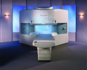 Most exams can be performed on the Open MRI such as brain, spine, pelvis and extremity studies. Although the resolution of some studies is not as good as high field MRI, this technology has become an alternative for claustrophobic patients who can not tolerate the high field magnet design. Imaging takes longer with this technology, usually one hour per exam.
Specialized neuroimaging studies, such as pituitary, optic nerve and internal auditory canal are best performed on a high field unit.
The MRI scan is performed inside a large cylindrical magnet with a patient table in the center. Conscious sedation is available at all locations for the highly claustrophobic patient. However, a doctor’s order is necessary and the appointment must be scheduled with conscious sedation so that the patient can be given special instructions prior to arrival at the MRI facility.
What to Expect:
You will be asked to fill out a questionnaire about your symptoms, prior studies and prior surgeries.
Because you will be entering a room with a strong magnet, we need to know of any metal or implants on or in your body. For example, pacemakers, metal implants, aneurysm clips, surgical staples, bullet wound or shrapnel, implanted drug infusion devise, bone stimulators, or permanent eyeliner. If you were issued a card with the exact manufacturer and serial number of the device, please bring that card with you to your appointment. If you have had occupational or recreational exposure to metal (welding, grinding, etc), you will need to have an radiograph to make sure that you don’t have any metal fragments in the eyes, because they can sometimes migrate in a strong magnet, even years after the exposure.
You will be asked to lie flat on the table, and then a “coil” will be placed over the area of interest. The coil typically looks like an athletic ice wrap and can be rigid or somewhat flexible. The table moves slowly into the scanner that houses a large magnet. During the procedure, you will be able to communicate with the technologist by an intercom. The technologist can also watch you through a small camera. The technologist will be in constant dialog with you and notify you when to expect a loud or unusual sound.
Typical open MRI studies require 60 minutes. It is important to remain motionless throughout the exam.
Some MRI exams require an injection of an MRI contrast or dye. This is completely different from the contrast agent or dye used for x-ray tests such as an IVP or CT scan. If you are over age 65 or have diabetes, high blood pressure or any history of kidney or severe liver disease or transplant we will need the results of blood tests for kidney function before we give intravenous contrast.
Preparation:
If you can, please leave loose jewelry and watches at home. Otherwise, you will be required to place them in a locker with your cell phone, glasses, and anything else that contains metal.
Magnetic Resonance Cholangiopancreatography (MRCP)
Nothing to eat or drink four hours prior to exam.
All other MR exams
There is no special preparation.
Nuclear Medicine
 Nuclear medicine studies provide information about the function of specific organs. Red blood cells or small proteins are tagged with a special tracer, and the machine records how the tracer is carried through or metabolized by the body. Usually, studies are performed to diagnose a disease or determine if the organ is functioning properly. In some cases, radiation is used to treat a condition, such as an overactive thyroid or thyroid cancer.
What is Radiation?
Radiation is a type of energy which comes from both natural and man-made sources. Examples of natural radiation include light and heat. Man-made radiation is used daily in microwave ovens as well as radio waves. Everyone is exposed to natural background ionizing which comes from outer space, the sun, the earth, the air, our food and drink, and from building materials such as concrete, bricks and mortar.
Is Nuclear Medicine Safe?
Nuclear medicine is safe because the radioactive tracers, or radiopharmaceuticals, used are quickly eliminated from the body in the urine or stool. The dose of radiation is very small and the tracers lose their activity very quickly. The amount of radiation exposure is typically less than a bicoastal airplane trip. Airline personnel can get as much radiation exposure as our patients get in all of their diagnostic tests combined.
Indications
Nuclear medicine scans can study any organ in the body, including the bloodstream. The most common tests are scans of the bones, heart, lungs, kidneys, gallbladder, and thyroid.
The purpose of these exams is to analyze kidney function, visualize heart blood flow and function, scan lungs for pulmonary embolism (blood clot), evaluate the gallbladder for inflammation (cholecystitis) or poor function, evaluate bones for fractures, infection, arthritis and tumors, determine the presence or spread of cancer in various parts of the body, identify location of bleeding in the bowel confirm and identify the location of infection, measure thyroid function in patients with symptoms of overactive or under active thyroid, and localize abnormal lymph nodes before surgery in patients with breast cancer or melanoma.
What to Expect:
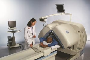
Depending on the scan ordered, the radiopharmaceutical may be given through an injection into the vein, swallowed, or inhaled through a nebulizer. The radiopharmaceutical will eventually concentrate in the organ of interest. A special camera called a gamma detector will be positioned close to the part of the body that is being scanned. The information is sent to a computer which processes and analyzes the amount and distribution of radiopharmaceutical in your body. The computer can display reconstructions or measurements. Depending on the test, the scan may be performed immediately or after a few days. Some scans may require multiple visits.
The radiopharmaceutical will naturally lose its radioactivity over time. It may pass out of your body through your urine or stool during the first few hours or days following the test. You may be instructed to take special precautions after urinating, to flush the toilet twice and to wash your hands thoroughly. You should also drink plenty of water.
Preparation:
You may be asked to switch into a gown for the procedure. You should inform the technologist of all medications that you are taking, including vitamins and herbal supplements. They will ask you about recent surgeries, medical conditions, and history of cancer. You should tell the technologist if you have any allergies. Also, let them know if you have diabetes or have had a blood test that detected abnormal kidney function at any time in your past.
You should always inform the technologist about any possibility that you are pregnant or if you are breastfeeding. They will give you special instructions for these two conditions. Jewelry and any other items with metal should be left at home since they can interfere with the study. You will be offered a locker to store personal items prior to the exam.
You will receive specific instructions on the day of your scan. If you are having a radioactive iodine (I-131) treatment, you will be given advance instruction and will meet with the radiologist on the day of the exam.
Locations for Nuclear Medicine:
- Memorial Regional Medical Center
- Richmond Community Hospital
- St. Francis Medical Center
- St. Mary’s Hospital
Positron Emission Tomography/Computed Tomography (PET/CT)
 Positron Emission Tomography in combination with Computed Tomography (PET/CT) provides a more accurate evaluation for cancer than either modality alone. CT provides the roadmap of precise anatomic detail (size and location of the tumor, mass, etc.), and a PET scan overlay identifies abnormal metabolic activity (cellular activity of the tumor, mass, etc.).
Anatomic
CT scanners send a small column of x-rays through the body, which are measured by detectors in the CT scanner. A computer algorithm processes the information to produce pictures of the body’s internal structures.
Metabolic
PET images take advantage of the fact that metabolically active organs and rapidly growing tumors consume glucose (sugar) at high rates. Fluorodeoxyglucose (FDG) a sugar similar to glucose is tagged with the radioactive fluorine (F18). As the FDG is used, it emits positrons. These positrons collide with electrons, giving off gamma rays, and a computer converts the gamma rays into images. These images demonstrate metabolic “hot spots.”
Large studies have shown that the following cancer types benefit most from this technology: breast, cervical, ovarian, esophageal, colorectal, lung, head and neck, sarcoma, melanoma and lymphoma.
PET/CT can be used to determine the extent of disease or the location of disease for biopsy, surgery or treatment planning. Using both modalities together enables the radiologist to make a more precise interpretation at an earlier time point. Later, it can be used to assess the response to treatment and detect residual or recurrent disease at an earlier time point. In this way, the patient may avoid additional biopsies.
What to Expect:
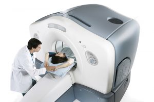
The entire examination usually takes less than 30 minutes. The tagged FDG solution is injected into a vein. Images are then taken in a machine that looks like a traditional CT machine, in which a table slides into a detector that looks like a large donut. It is important that the patient remain still during the test since the two images must be superimposed and the detection of subcentimeter disease is a desired benefit of this technology.
Preparation:
No preparation is necessary
Locations for PET/CT:
- Memorial Regional Medical Center
- St. Francis Medical Center
- St. Mary’s Hospital
Ultrasound
Ultrasound uses high frequency sound waves to create still or video images of the inside of the body. There is no ionizing radiation used, so it is safe for pregnant women and children. Pictures are obtained by placing gel on the skin and moving an ultrasound transducer over the area of interest. The exam is usually painless.
Fetal or obstetrical ultrasound is most often used to evaluate the size and age of a fetus as well as assess the growth during pregnancy. It can also be used to screen for certain abnormalities fetal anatomy.
Abdominal ultrasound evaluates the gallbladder, liver, spleen, pancreas, and spleen. Size and shape of the organs are usually easily recognized. Abnormalities, such as gallstones or kidney cysts, can be identified using ultrasound.
Pelvic ultrasound. In women, detailed images are obtained of the uterus and ovaries. This study usually includes an endovaginal exam, which is performed using a specially shaped transducer covered by a sterile sheath that the patient is asked to place into her vagina. In men, pelvic ultrasound can be utilized to image the prostate gland. The overall size and shape of the gland can be estimated. Prostate nodules can be imaged using an endorectal approach.
Thyroid ultrasound assesses the size of the thyroid gland. Thyroid lesions and/or nodules can be characterized for their make-up and location.
Vascular ultrasound is used to assess arteries and veins. Pulsed Doppler and color flow Doppler are used in addition to standard ultrasound imaging. Veins (usually leg veins) can be evaluated for thrombosis (blood clot) and arteries can be scanned to determine if there is any narrowing of the vessel due to atherosclerosis, usually in the carotid arteries of the neck. Vascular ultrasound can also be used to evaluate the abdominal blood vessels.
Breast ultrasound is performed to evaluate a palpable lump or an abnormality visualized on mammography or MRI. The distinction can be made between fluid-filled cysts, lymph nodes and solid masses. This information helps the radiologist and the patient’s clinician determine the appropriate course of treatment for the patient.
Musculoskeletal ultrasound is a targeted exam to evaluate individual tendons for small tears that cannot be seen on MRI.
Preparation:
- Pelvic and Obstetrical (up to 20 weeks) Drink 4 (8 oz.) glasses of liquid without carbonation (no soda) one hour before your appointment. Be sure to eat. Do not chew gum. Do not empty your bladder. Obstetrical patients (20 weeks to term) drink 1 (8 oz) glass only.
- Abdominal (Gallbladder, Liver, Pancreas, RUQ, Spleen) DO NOT eat or drink after midnight before your exam. Do not chew gum.
- Breast, Thyroid, Musculoskeletal, and Vascular, no preparation necessary
Locations for Ultrasound:
- Memorial Regional Medical Center
- Richmond Community Hospital
- St. Francis Medical Center
- St. Mary’s Hospital
- Bon Secours Imaging at Innsbrook
- Bon Secours Imaging at Reynolds
- Bon Secours Imaging at Short Pump
- Bon Secours Imaging at Westchester
- Laburnum Diagnostic Imaging Center (breast ultrasound only)
- St Mary’s Women’s Imaging Center (breast ultrasound only)
X-rays (General Radiology)
Digital radiographs are the most common examination performed in our department. A small column of radiation passes through the body and strikes the digital sensor on the other side. As with digital photography, the image can be adjusted. The radiologist can magnify, rotate, lighten/darken, or measure on a workstation to better answer the clinical question. Radiographs of the chest, abdomen, spine, sinuses and extremities are the most common tests performed.
What to expect:
At least two images are taken; additional images may be needed to answer the clinical question. You may be asked to wait, if your physician has requested that the result be called to their office prior to your departure from the center.
Preparation:
There is no special preparation. You may need to change into a gown upon arrival. Typically, these studies require 5 to 15 minutes. Radiographs are offered on a walk-in basis, with no appointment necessary. If you are pregnant, you will need to notify the technologist.
Locations for X-rays:
- Memorial Regional Medical Center
- Richmond Community Hospital
- St. Francis Medical Center
- St. Mary’s Hospital
- Bon Secours Imaging at Innsbrook
- Bon Secours Imaging at Reynolds
- Bon Secours Imaging at Short Pump
- Bon Secours Imaging at Westchester